What are the risk points and pitfalls of femoral condyle fracture?
Supracondylar femur fractures presentunique challenges for orthopedic surgeons, particularly in the context of anaging population and increasingly active lifestyles. This article will delveinto the pitfalls and danger points associated with these fractures, armingyoung orthopedic surgeons with the knowledge needed to avoid complications andachieve optimal patient outcomes.
Understanding the Complexity
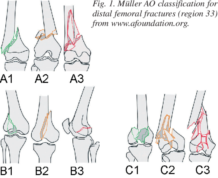
Supracondylar femur fractures are fracturesoccurring in the distal femur just above the knee joint. They often result fromhigh-energy trauma in younger individuals and low-energy falls in the elderlywith osteoporotic bone. These fractures can be complex, involving articularsurfaces, significant comminution, and soft tissue injuries.
Missed Associated Injuries: A CriticalFirst Step
A thorough initial assessment is crucial.Always remember that supracondylar femur fractures seldom occur in isolation.Be vigilant for:
· Femoral neck fractures: Thesecan be subtle and easily missed. A high index of suspicion is warranted,especially in elderly patients.
· Patellar fractures: Directtrauma or forceful quadriceps contraction can lead to patellar fractures.
· Tibial plateau fractures:
Axial loading forces can transmit tothe tibia, causing plateau fractures.
Missing any of these associated injuriescan lead to inadequate treatment, prolonged pain, and functional limitations.Employ a systematic examination, supplemented by appropriate imaging studies(X-rays, CT scan) to ensure no injury is overlooked.
The Looming Threat of Vascular Injury
The proximity of the popliteal artery tothe distal femur makes it vulnerable to injury. Always assess vascular statusmeticulously, evaluating for:
· Diminished or absent pulses:
Compare the affected limb to thecontralateral side.
· Abnormal capillary refill:
Delayed refill suggests compromisedcirculation.
· Sensory changes:
Numbnessor tingling can indicate nerve compression from a hematoma or direct injury.
· Coolness and pallor: Signsof decreased perfusion.
If vascular compromise is suspected,immediate action is imperative
Obtain a CT angiogram to delineate theinjury and consult a vascular surgeon promptly for potential surgicalintervention. Delay can lead to irreversible limb ischemia and amputation.
Compartment Syndrome: A SurgicalEmergency
Distal femur fractures, especiallyhigh-energy injuries, carry a risk of compartment syndrome. Early recognitionis essential:
· Pain: Out of proportion tothe injury and unrelieved by analgesics.
· Pallor: Pale skin due todecreased blood flow.
· Paresthesia: Numbness ortingling.
· Pulselessness: A late andominous sign.
· Paralysis: Loss of musclefunction, indicating severe ischemia.
Don't rely solely on one sign. Regularlyre-evaluate the patient for these symptoms and maintain a low threshold formeasuring compartment pressures.
Promptfasciotomy is the definitivetreatment for compartment syndrome, relieving pressure and restoring bloodflow. Delay can result in muscle necrosis, permanent nerve damage, andfunctional impairment.
Inadequate Articular Reduction:Long-Term Consequences
The articular surface of the distal femurplays a critical role in knee joint function. Failure to achieve anatomicalreduction can lead to:
· Post-traumatic arthritis:
Joint incongruity accelerates wear andtear, causing pain and stiffness.
· Functional limitations:
Malalignmentcan restrict range of motion and impair activities like walking, running, andstair climbing.
Precise reduction is paramount
Utilize appropriate techniques,including:
· Traction:
Gently pullingon the distal fragment to restore length and alignment.
轻轻拉动远端碎片以恢复长度和对齐。
· Joysticks:
Instrumentsthat allow controlled manipulation of fragments.
· Clamps:
Holding reducedfragments in place before definitive fixation.
Intraoperative fluoroscopy is indispensablefor confirming satisfactory reduction.
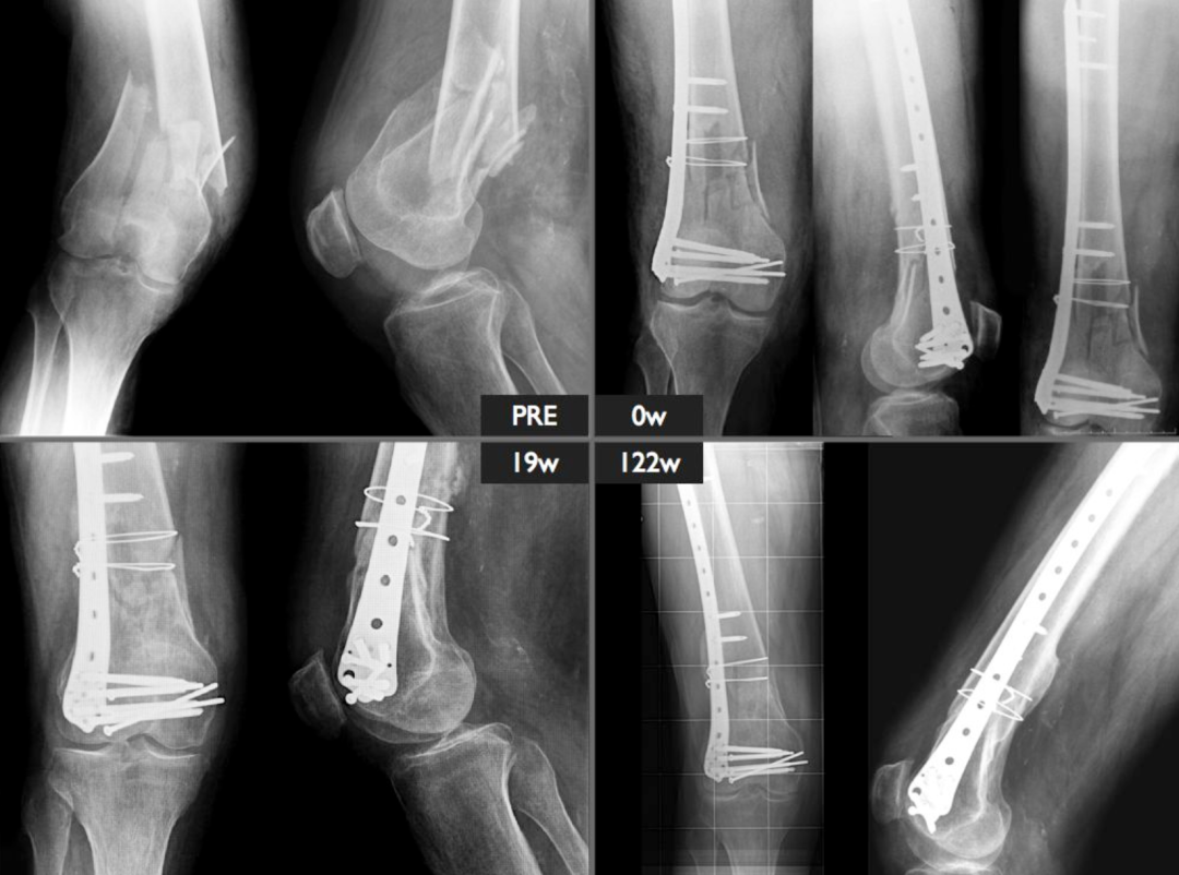
Retrograde Intramedullary Nailing: NotWithout Its Perils
Retrograde intramedullary nailing is acommon technique for stabilizing distal femur fractures. However, be mindful ofits limitations and potential complications:
· Subtrochanteric Fractures:
Retrograde nailing is generallycontraindicated, as adequate proximal fixation is difficult to achieve.
· Limited Knee Mobility:
Preexistingknee stiffness or limited range of motion can hinder nail insertion.
· Posterior Guidewire Placement:
Erroneous guidewire placement andreaming in the posterior intercondylar notch can injure the posterior cruciateligament.
· Reamer/Nail Impingement:
The distal end of the nail must be placed deep to thearticular surface to prevent impingement on the patella. Ensure adequatecountersinking.
Meticulous surgical technique and carefulattention to anatomical landmarks are essential for safe and effectiveretrograde nailing.
Postoperative Care: A Vital Bridge toRecovery
Postoperative management is equally crucialfor optimizing outcomes:
· Early mobilization:
Encourageearly range of motion exercises to prevent stiffness and promote circulation.
· Weight-bearing precautions:
Follow weight-bearing restrictions asdetermined by fracture stability and fixation construct.
· Pain management:
Provideadequate pain control to facilitate early mobilization and rehabilitation.
· Regular follow-up:
Monitorhealing progress with radiographs, assess for complications, and adjust thetreatment plan as needed.
Conclusion
Supracondylar femur fractures demand anuanced approach, combining thorough assessment, meticulous surgical technique,and attentive postoperative care. By understanding the potential pitfalls anddanger points outlined in this article, young orthopedic surgeons canconfidently navigate the complexities of these fractures, minimizingcomplications, and maximizing patient outcomes.